![]() Table 3 of
Dansault, Mol Vis 2007;
13:511-523.
Table 3 of
Dansault, Mol Vis 2007;
13:511-523.
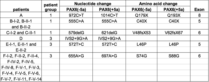
Table 3. Summary of detected mutations and changes
We show the pedigrees of the affected patients whenever they were available with certainty. We also indicate the sequence of the six mutations reported in this study. Each mutation is associated with the affected patients from the clinical group 1 corresponding to aniridia phenotypes or from the group 3 corresponding to other ocular phenotypes associated in one large family (F) with neurological anomalies. A and D are isolated cases whereas B, C, E, and F refer to familial cases (see Figure 2). The exon in which the mutated amino acid residue is located is mentioned. Each mutation is named according to the nucleotide change and the corresponding amino acid modification for the two major isoforms: Pax6(-5a), comprising 422 amino acids, and PAX6(+5a), comprising 436 amino acids resulting from alternative splicing.