![]() Figure 7 of
Biswas, Mol Vis 2007;
13:345-359.
Figure 7 of
Biswas, Mol Vis 2007;
13:345-359.
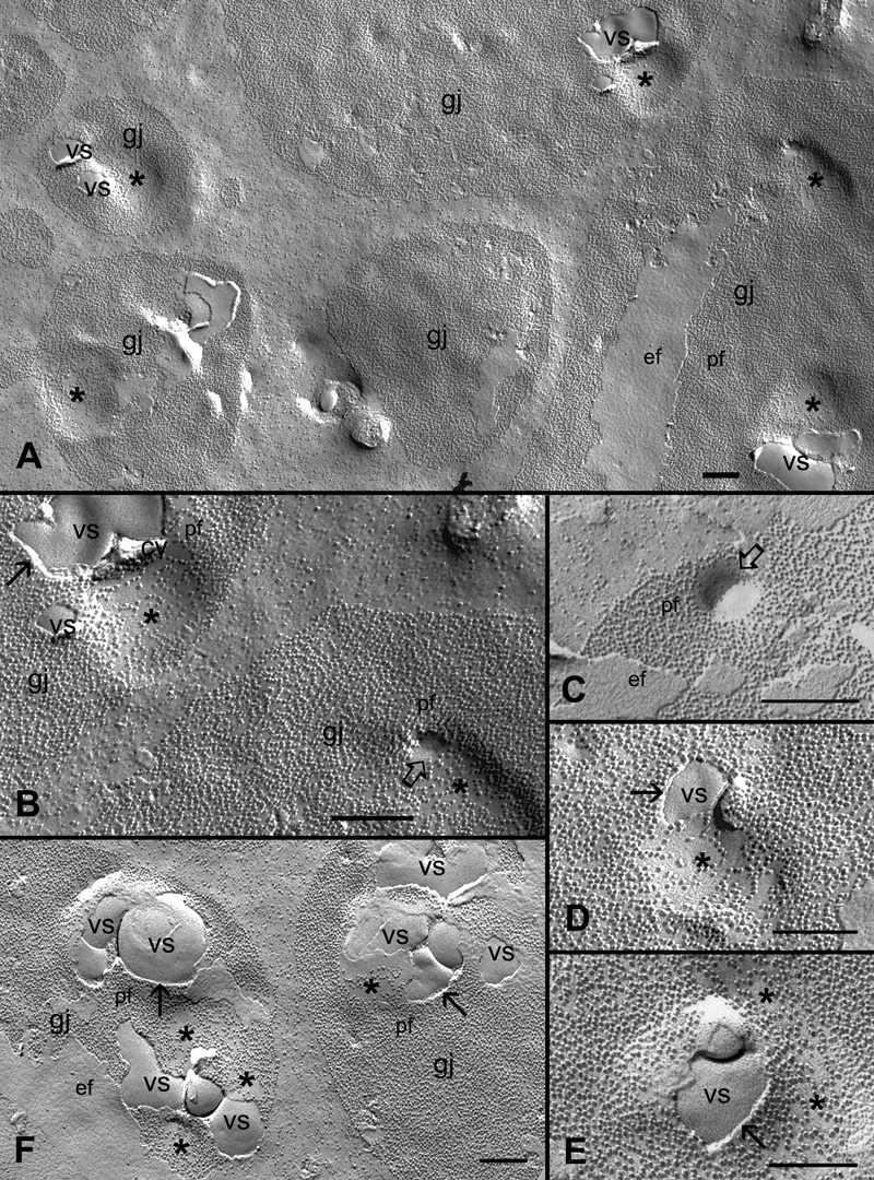
Figure 7. Specific association of unique vesicular structures with gap junction plaques in the outer cortical fibers
A: A low magnification shows a number of vesicular structures (vs) specifically associated with gap junction plaques (gj) in the outer cortical fibers. Asterisks denote the presence of small concaved connexon particles-less or -free areas often associated with the vesicular structures. B and C show the indication of endocytosis (open arrow) of the P-face (pf) of the connexon-free membrane within the gap junction plaques. D and E exhibit the close association of vesicular structures with the adjacent connexon-less membrane areas (asterisk). F: A cluster of vesicular structures with different shapes and sizes are accumulated in two adjacent gap junction plaques. The distinct separation between the P-face of the junction membrane and the vesicular structures is indicated by the arrows. These vesicular structures are shown to be enriched with cholesterol (see Figure 9). The scale bars are equal to 200 nm.