![]() Figure 9 of
Biswas, Mol Vis 2007;
13:345-359.
Figure 9 of
Biswas, Mol Vis 2007;
13:345-359.
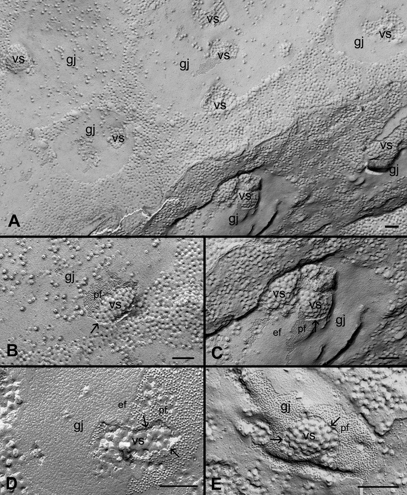
Figure 9. Filipin-treated experiments reveal the presence of discrete filipin-cholesterol particles specifically associated with individual cholesterol vesicles (vs) in the different subtypes of cholesterol-containing gap junctions (gj)
A: A low magnification shows a number of cholesterol vesicles distributed randomly within the gap junction plaques. B through E Higher magnifications illustrate the specific accumulation of filipin-cholesterol particles in the vesicular structures (vs) of various forms. The arrows indicate the distinct separation of individual cholesterol vesicles from the gap junction membranes. The scale bars in A through C are equal to 200 nm and in D and E are equal to 100 nm.