![]() Figure 5 of
Zhang, Mol Vis 2007;
13:2289-2300.
Figure 5 of
Zhang, Mol Vis 2007;
13:2289-2300.
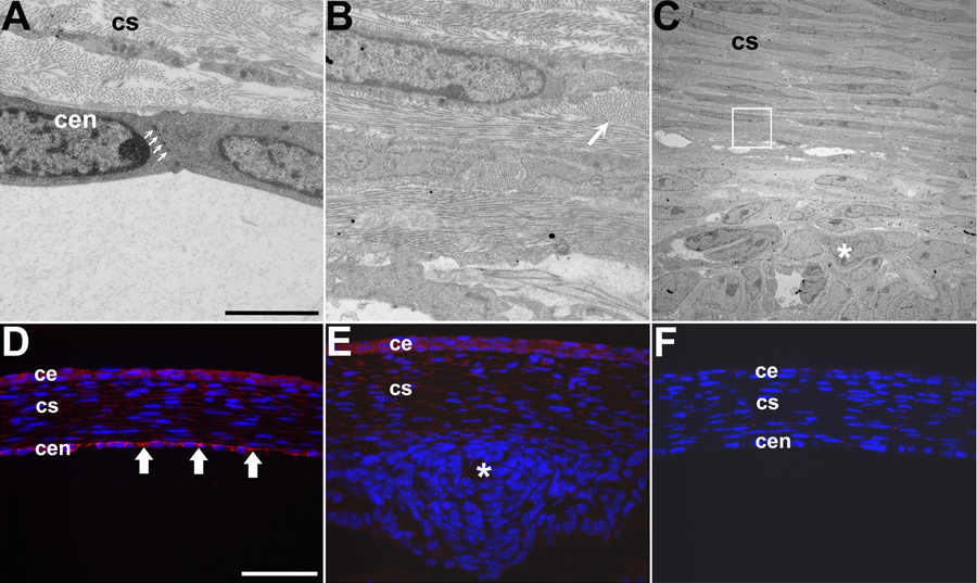
Figure 5. Altered corneal endothelial differentiation
Electron micrographs of P2 nontransgenic (A) and DREAM-Tox176 (OVE1757; B,C) corneal endothelium and immunohistochemistry for Z01 (D-F). B is a higher magnification of the boxed portion in C. Cells in the corneal endothelium are normally connected by tight and adherens junctions (A, arrows). These junctions were not detected in the Tox176 transgenic corneas. Collagenous material (B, arrows) was seen between and around all corneal cells. Z01 expression was seen in the nontransgenic corneal endothelial cells (D, arrows), but was not detected in the inner cells of the Tox176 cornea (E). The Z01 antibody was left out for the section shown in F. The asterisks in C and E indicate abnormally migrating iridial mesenchymal cells (see Figure 6 H and X). Abbreviations; ce, corneal epithelium; cen, corneal endothelium; cs, corneal stroma. Scale bar in A applies to A, B, and C. Scale bar in D applies to D, E, and F. Scale bar (C) represents 1,250 nm in A and B and 10 μm in C. Scale bar (D) represents 50 μm in D-F.