![]() Figure 6 of
Zhang, Mol Vis 2007;
13:2289-2300.
Figure 6 of
Zhang, Mol Vis 2007;
13:2289-2300.
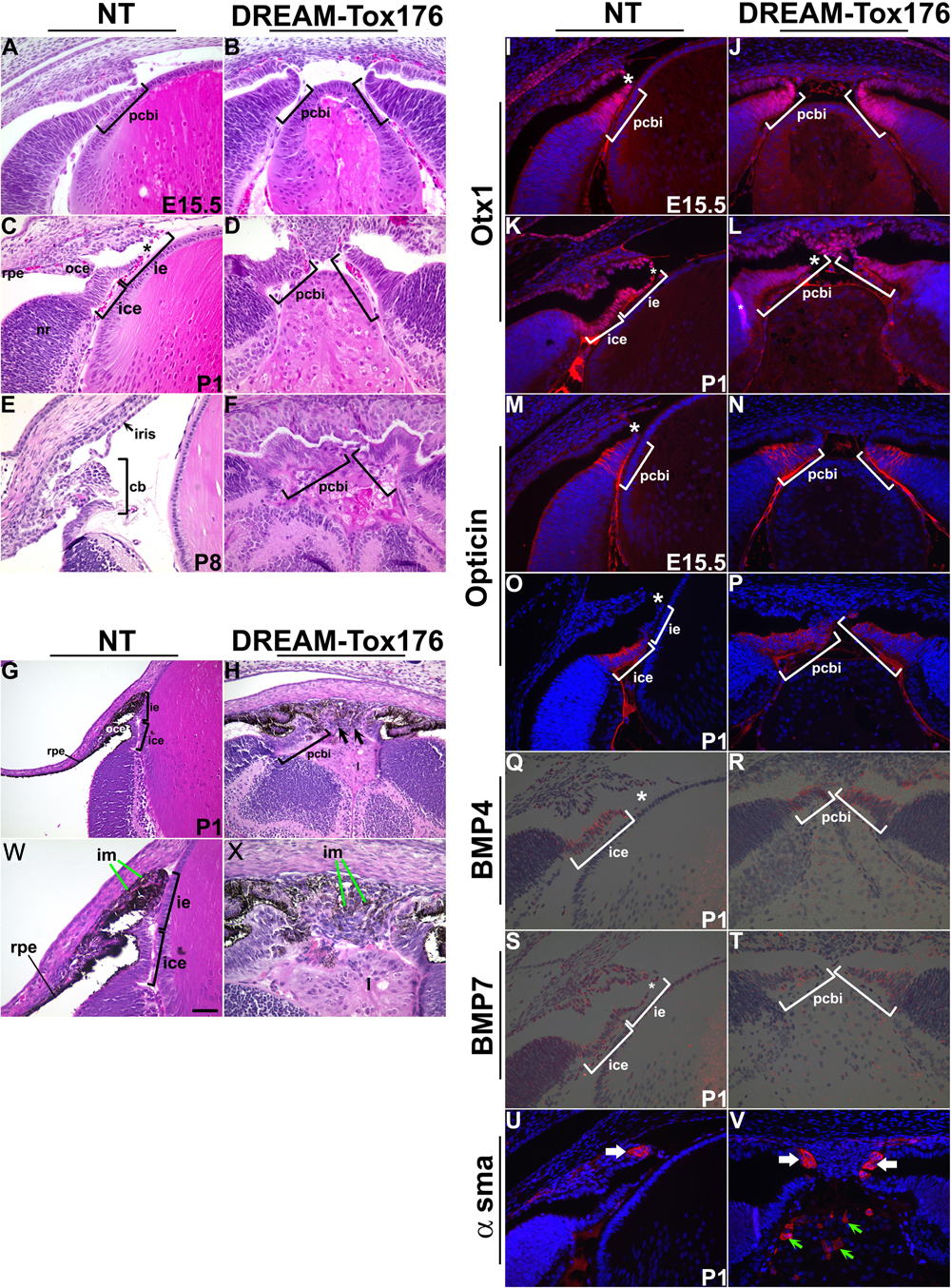
Figure 6. Iris and ciliary body defects
A-H,W,X: Hematoxylin and eosin staining of ocular sections of E15.5, P1 and P8 nontransgenic (NT) and DREAM-Tox176 mice. At P1, the iris epithelium (ie) can be distinguished in the nontransgenic (C) but not in the Tox176 transgenic eyes (D). Ciliary folds are indicated by brackets in E and F. Ocular sections of pigmented newborn offspring are shown in G-H. W and X are higher magnifications of panels G and H, respectively. Pigmentation can be seen in the retinal pigmented epithelium (RPE), outer ciliary epithelium (Oce; G) and in cells of the presumptive iris (im, X). Immunohistochemistry on E15.5 and P1 ocular sections to detect expression of Otx1 (I-L), opticin (M-P), and smooth muscle actin (α-sma). The antibody staining is in red and the blue DAPI stain marks the nuclei. Nontransgenic iris epithelial cells do not express opticin (O), but all cells of the Tox176 presumptive ciliary body and iris express opticin (P). Expression of α-smooth muscle actin in the putative iris sphincter muscle is unaltered (U,V, white arrows). Expression of α-smooth muscle actin was also seen in the fibrotic cells of the lens (V, green arrows). In situ hybridizations were performed on P1 ocular sections to assay for expression of BMP4 (Q,R) and BMP7 (S,T). Abbreviations; ice, inner ciliary epithelium; l, lens; nr, neuroretina; pcbi, presumptive ciliary body and iris. Discontinuities within the presumptive ciliary body and iris are artifacts of the histological processing and are indicated as asterisks in C, I, K, L, M, O, Q, and S. Scale bar (W) represents 50 μm in A-V and 25 μm in W and X.