![]() Figure 3 of
Okamoto, Mol Vis 2007;
13:2112-2118.
Figure 3 of
Okamoto, Mol Vis 2007;
13:2112-2118.
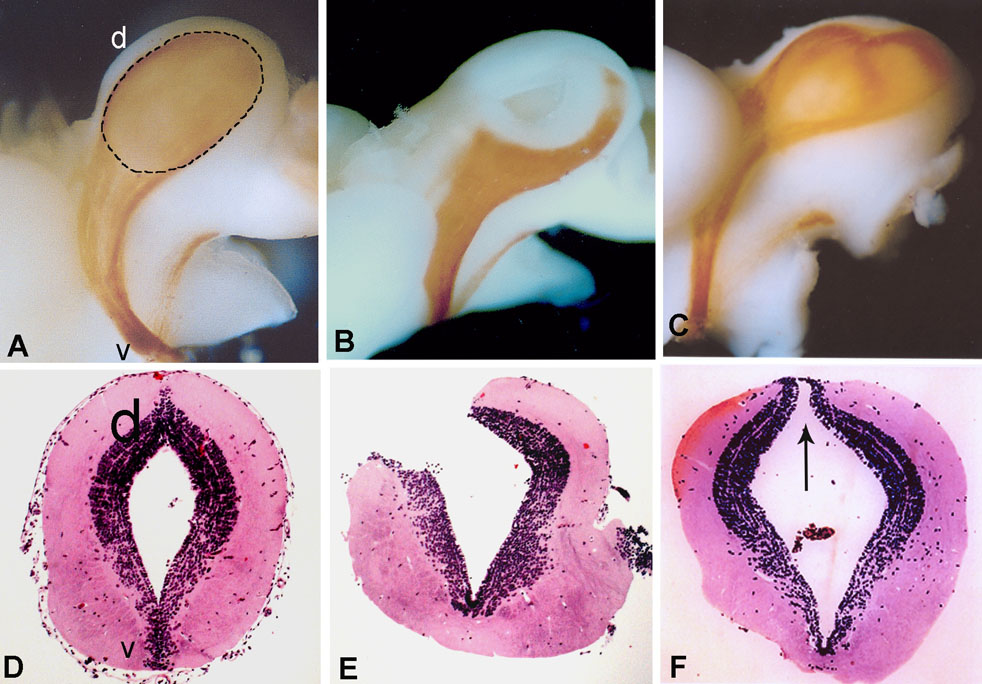
Figure 3. Regeneration of the retinotectal projections
Regeneration of the retinotectal projections pattern after excision of the left optic tectum (OT) of the midbrain (mesencephalon). A-C: Visualization of the retinotectal pattern in the midbrain and interbrain (diencephalon) by horseradish peroxidase staining of optic nerve fibers. A, Un-operated left OT showing the normal patterns of labeled retinotectal projections. Dotted line defines the OT. B, Left OT just after excision with apparent loss of the retinotectal projections due to the operation. C: Regenerating left OT six months after operation, with recovery of the retinotectal projections, as indicated by the staining with HRP. Compare the similarity in the staining patterns with A. D-F: Shown are hematoxylin and eosin-stained transverse sections of the midbrain that correspond to figures in A-C, respectively. D: Unoperated midbrain. E: A cross section through the excised tectum (corresponding to B) showing a missing large area due to the removal of the tectum tissue. F: A cross section through a regenerating tectum (corresponding to C) six months after excision. Note that the tissue has been regenerated and that it looks considerably similar to un-operated brain shown in D. Arrow indicates the remnant of the wound in this sample. The top part of the tissue in A-C and of the sections in D-F is the dorsal (d) and the bottom is the ventral (v) side of the midbrain. The minor tract of labeled axons outside the left optic tectum is the tractus opticus accessory [15]. These tracts are located at the interbrain region. According to the orientation of the specimens in A-C the endbrain is located on the left of the stained tracts and the hindbrain on the right (see also illustration in Figure 1).