![]() Figure 1 of
Okamoto, Mol Vis 2007;
13:2112-2118.
Figure 1 of
Okamoto, Mol Vis 2007;
13:2112-2118.
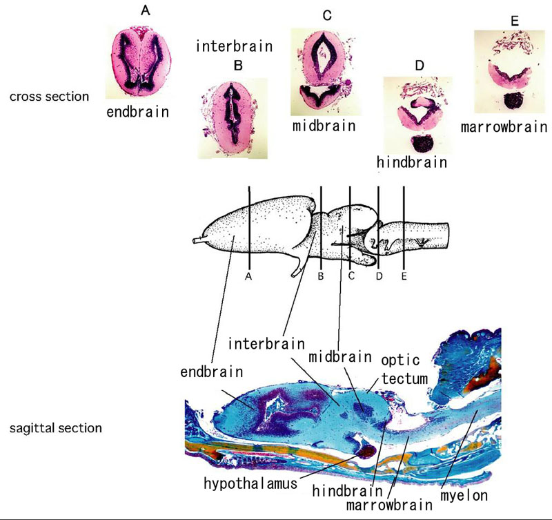
Figure 1. Histological sections and anatomy of the newt brain
This figure shows the basic anatomy of the newt brain and aims to orient the reader in the sections of the brain that were analyzed. In the middle of the panel we can see the overall anatomy of the newt brain. On the top there are cross sections (A-E), which correspond to the different parts of the brain (corresponding to lines A-E in the middle of the panel). On the bottom there is a sagittal section showing also the corresponding parts. A: endbrain, B: interbrain, C: midbrain, D: hindbrain, E: marrowbrain.