![]() Figure 7 of
Wajchman, Mol Vis 2007;
13:1902-1911.
Figure 7 of
Wajchman, Mol Vis 2007;
13:1902-1911.
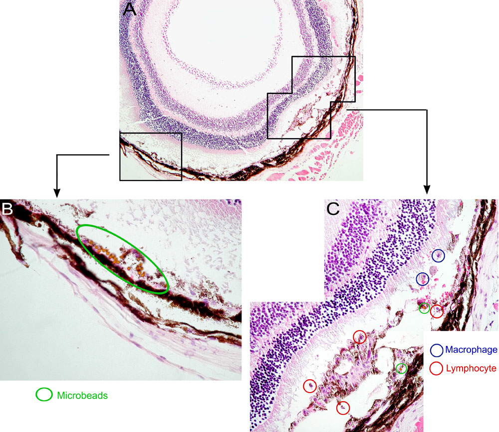
Figure 7. Early inflammation in transplanted OT1 mice
In a separate experiment, OT1 mice were injected with retinal pigment epithelium (RPE) and microbeads as described in the legend for Figure 6. A larger section of the eye is shown (A) at 40X magnification with two sections outlined in black, which are shown at a higher magnification (400X; B) and (C). Examples of macrophages, large cells with pericentric nuclei and abundant cytoplasm, are circled in blue whereas examples of lymphocytes, with large nuclei and only a rim of cytoplasm are circled in red. In contrast, much more abundant numbers of lymphocytes are detectable in OT1 recipients at week two after transplantation of TRP-1-OVA RPE (the lower right panel of Figure 6).