![]() Figure 6 of
Wajchman, Mol Vis 2007;
13:1902-1911.
Figure 6 of
Wajchman, Mol Vis 2007;
13:1902-1911.
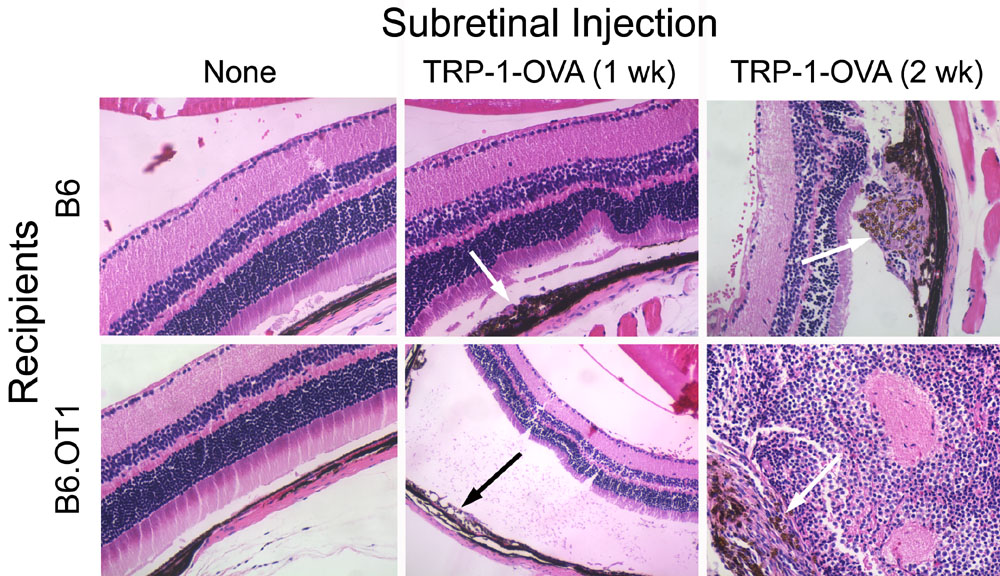
Figure 6. Ovalbumin as a neo-transplantation antigen
Retinal pigment epithelium (105) from B6 TRP-1-OVA mice were mixed with (105) microbeads and injected into the subretinal space of B6 or OT1 mice. Eyes from these mice and uninjected control mice were sectioned and stained with H&E; images were made at 200X except for the middle panel on the lower row, which is shown at 100X. Representative slides are shown to illustrate the retinal morphology and to detect leukocytic infiltration from at least 10 sections/eye and from two or three mice/group that were examined in this experiment. Arrows point to transplanted RPE and microbeads in the SRS.