![]() Figure 3 of
Giordano, Mol Vis 2007;
13:1842-1850.
Figure 3 of
Giordano, Mol Vis 2007;
13:1842-1850.
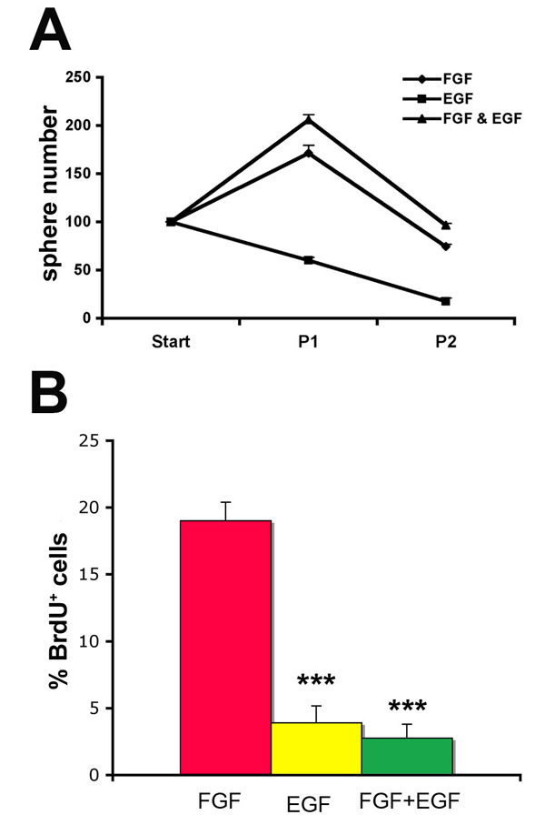
Figure 3. In vitro passaging of retinal spheres
A: Spheres were counted after they had been split one (P1) or two (P2) times. Start indicates the primary culture. Treatment with fibroblast growth factor (FGF) doubled the number of spheres recovered after the first passage. B: Shown are the percentages of proliferating, bromodeoxyuridine (BrdU) labeled cells (BrdU+), in retinal spheres cultured for seven days after the first passage (P1) in vitro. Cell proliferation after the first passage remained similar to primary cultures (see Figure 1D, D7). The Student t-test was used, and significance was calculated by comparing spheres grown with FGF to spheres grown with either epidermal growth factor (EGF) or FGF+EGF. Triple asterisk (***) indicates p<0.001.