![]() Figure 1 of
Giordano, Mol Vis 2007;
13:1842-1850.
Figure 1 of
Giordano, Mol Vis 2007;
13:1842-1850.
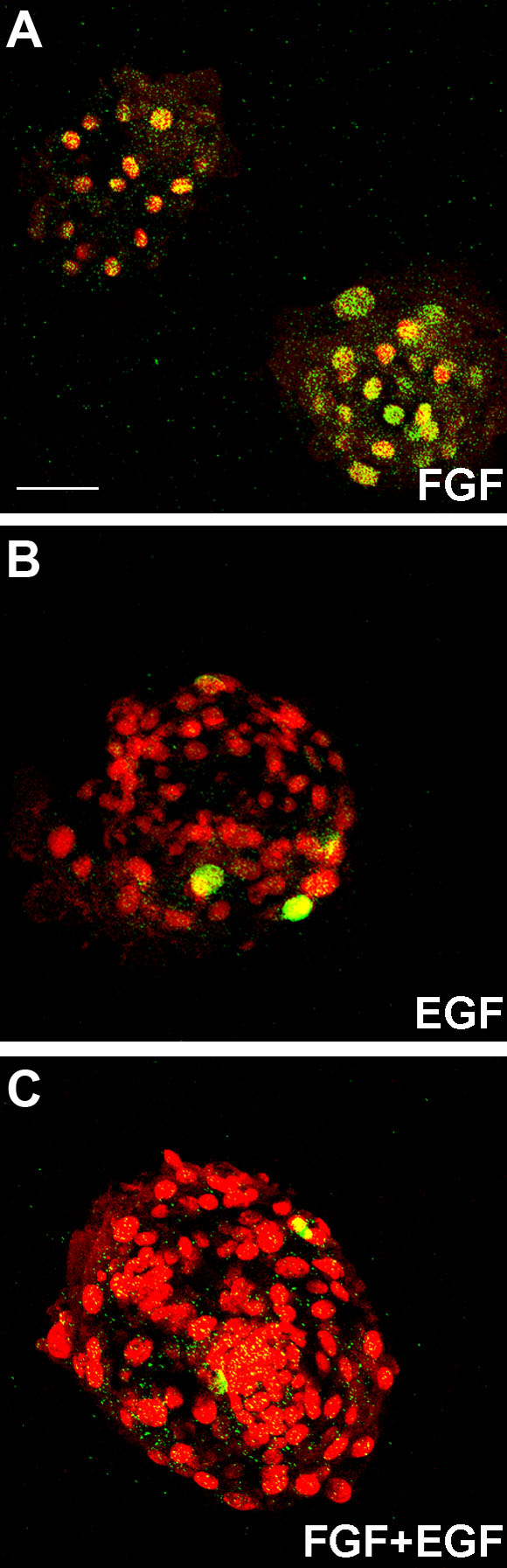
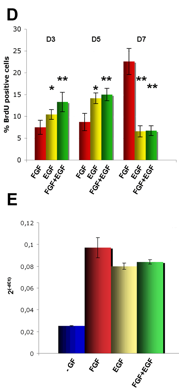
Figure 1. Proliferation of retinal spheres
A-C: Confocal images of bromodeoxyuridine (BrdU) staining (green) of retinal spheres cultured for seven days in the presence of either fibroblast growth factor (FGF; A) or epidermal growth factor (EGF; B), or FGF+EGF (C). Nuclei were stained with propidium iodine (red). There was a proliferation of cells at the seventh day of culture when retinal spheres were grown with FGF. Scale bar represents 60 μm. D: Percentage of BrdU+ cells counted in spheres at the third day, fifth day, or seventh day of culture (D3, D5, and D7). Values are shown as percentage of BrdU+ stained nuclei versus the total number of nuclei and are the average of BrdU+ cells counted in five spheres of three different experiments. The Student's t-test was performed. Asterisk (*) indicates p<0.05, and double asterisk (**) represents p<0.01. Significance was calculated by comparing spheres grown with FGF to spheres grown with either EGF, or FGF+EGF. E: Quantitative analysis of Cyclin D1 mRNA expression in spheres cultured for seven days. Spheres grown in the presence of (FGF, EGF or FGF+EGF) expressed a significant higher level of Cyclin D1 than grown in the absence of growth factor (-GF), but there was no significant measured difference among cultures with different GFs.