![]() Figure 6 of
Mott, Mol Vis 2007;
13:1802-1812.
Figure 6 of
Mott, Mol Vis 2007;
13:1802-1812.
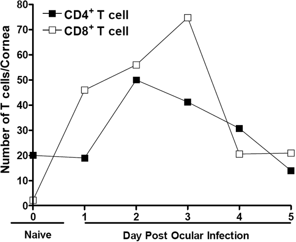
Figure 6. Cell sorting of CD4+ and CD8+ T cells in corneas of infected mice
BALB/c mice were ocularly infected with 2x10⁵ PFU/eye of HSV-1 strain McKrae as described in the legend to Figure 5. Corneas from 10 mice per time point were isolated without contamination from other parts of the eye as described in Methods. Ten naive mice were also used as control. Single cell suspensions of corneas were reacted with mAbs to 7-ADD/CD4/CD8, and the number of CD4+ or CD8+ T cells and 7-ADD- T cells were sorted using a MoFlo (Dako). Data are presented as the number of CD4+7-ADD- or CD8+7-ADD- cells per cornea.