![]() Figure 2 of
Hogewind, Mol Vis 2007;
13:1793-1801.
Figure 2 of
Hogewind, Mol Vis 2007;
13:1793-1801.
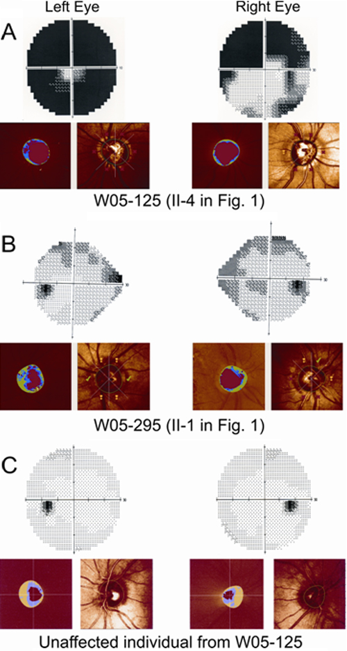
Figure 2. Phenotypic characterization of the probands with p.Phe430Leu mutation in the MYOC gene
Each panel has two parts; the upper part depicts the visual field printouts of the Humphrey Field Analyzer (HFA) and the lower part shows screenshots from Heidelberg Retina Tomograph II (HRT) analysis to detect loss of the papillary neuro-retinal rim. The right and left columns correspond to the right and left eyes of the probands, respectively. A: HFA and HRT results for proband of W05-125 (II-4 in Figure 1B) is shown. The large dark areas in the HFA results correspond to glaucomatous scotomas due to nerve fiber layer defects. HRT scans demonstrate glaucomatous increased optic disc cupping (red areas) and suspect (yellow exclamation marks) or manifest pathological (red crosses) neuro-retinal rim measures (according to the Moorfield's regression analysis) within the different quadrants of the optic disc. B: A similar representation as A is shown but for proband of W05-295 (II-1 in Figure 1C). The results are less severe than in proband W05-125, however, a nasal step characteristic for glaucoma can be observed in HFA, and a thinned neuro-retinal rim (blue-green area) can be seen by HRT. C: HFA and HRT results for a 55-year-old, unaffected individual from W05-125 are shown for comparison. HFA results indicate that there is no darkening due to glaucomatous defects on HFA. The small temporal black area corresponds to the physiological blind spot. HRT results show no thinning of the neuro-retinal rim is visible.