![]() Figure 16 of
Rizzolo, Mol Vis 2007;
13:1259-1273.
Figure 16 of
Rizzolo, Mol Vis 2007;
13:1259-1273.
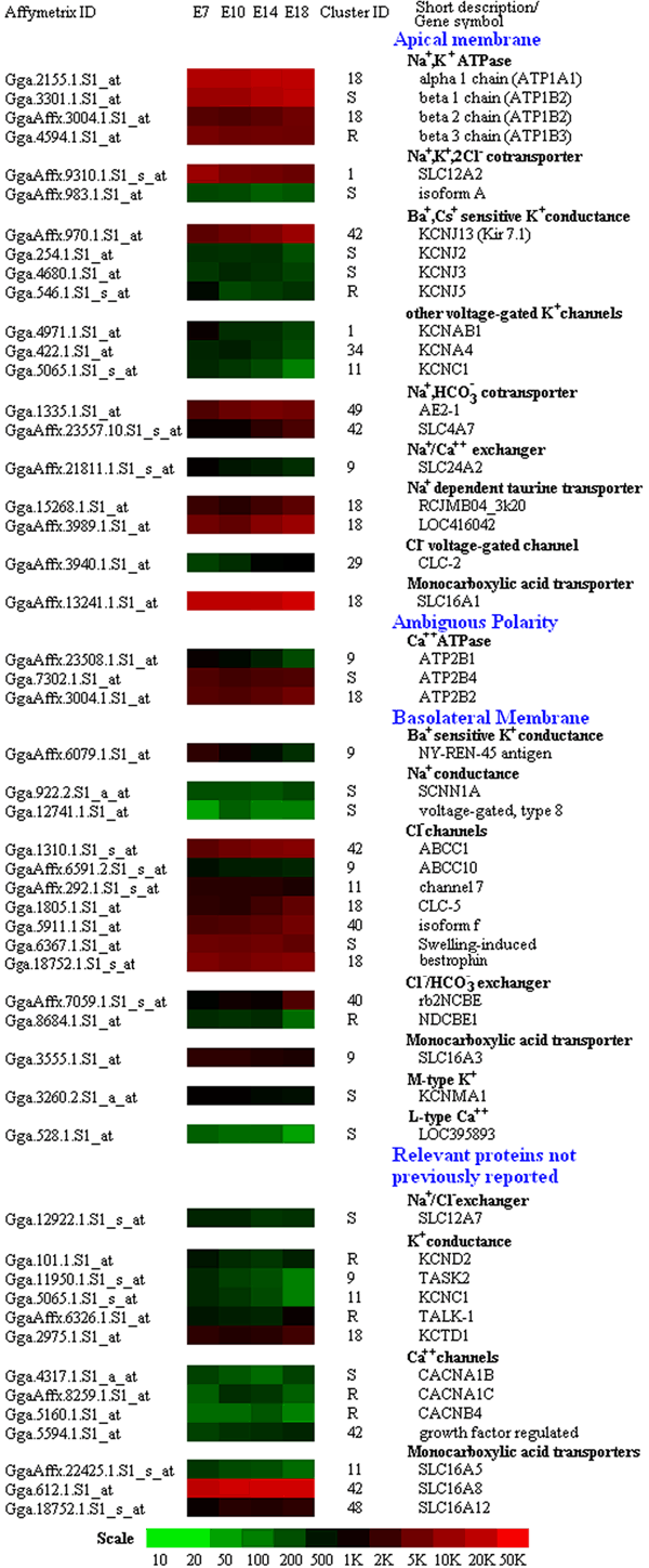
Figure 16. Ion transport protein genes expressed in retinal pigment epithelium
Transporters are grouped according to their subcellular location, as reported in recent reviews [72,73]. Additional transporters of the plasma membrane were also detected and are included at the bottom of the figure. For many of the transporters predicted by physiologists, a number of candidates were identified. Surprisingly, candidates with similar functions sometimes replaced one another during development. An example is the Ca++-ATPase, ATP2B1, mRNA decreased as the ATP2B2 mRNA increased. Mean values of 3-4 biological replicates are indicated according to the scale, in arbitrary units, depicted at the bottom. Complete gene titles and mean values with the associated p-statistic are listed in the corresponding Appendix 2.