![]() Figure 5 of
Lin, Mol Vis 2007;
13:1203-1214.
Figure 5 of
Lin, Mol Vis 2007;
13:1203-1214.
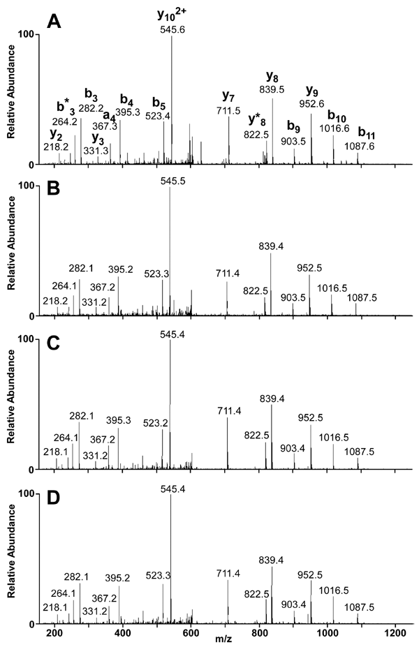
Figure 5. fragment ion spectrum spectra corresponding to the retinal G protein-coupled receptor splice isoform splice-site peptide
Protein bands, which correspond to the immunoreactive bands in the western blot of Figure 4, were stained with Coomassie blue, excised, and analyzed by liquid chromatography-mass spectrometry (LC/MS/MS). Fragment ion spectrum (MS/MS) spectra from precursor ions at m/z 613.4 matching the spectrum for the RGR-d splice-site peptide were observed for (A) recombinant RGR-d, (B) the about 24-kDa band from donor CA-066, (C) the about 55-kDa band from donor CA-066, and (D) the about 24-kDa band from donor O. For the spectra from the donor samples, background subtraction was used to eliminate low levels of constant chemical noise evident in the MS/MS spectra obtained at the precursor m/z, mass-to-charge ratio.
