![]() Figure 4 of
Lin, Mol Vis 2007;
13:1203-1214.
Figure 4 of
Lin, Mol Vis 2007;
13:1203-1214.
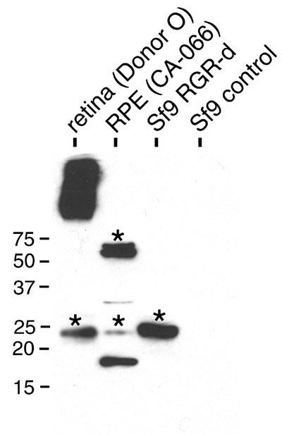
Figure 4. Western immunoblot assay of retinal G protein-coupled receptor splice isoform in the neural retina of donor O, retinal pigment epithelium of donor CA-066, and baculovirus-transduced Spodoptera frugiperda whole cell extract
Untreated Spodoptera frugiperda (Sf9) whole cell extract served as a negative control. The blot was probed with the DE17 antibody. The asterisks indicate protein bands that were cut from a parallel gel for liquid chromatography-mass spectrometry (LC/MS/MS) analysis. RPE represents retinal pigment epithelium.