![]() Figure 2 of
Mitchell, Mol Vis 2007;
13:1144-1153.
Figure 2 of
Mitchell, Mol Vis 2007;
13:1144-1153.
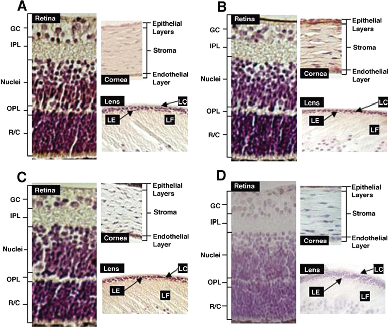
Figure 2. Immunocytochemistry analysis of the expression patterns of RhoA, Rac1, and Cdc42 in postnatal mouse eye
A: Expression of RhoA in different compartments. RhoA was highly expressed in the photoreceptors, the horizontal/amacrine/Muller cell layers, and in some ganglion cells. RhoA expression is relatively lower in other ganglion cells and plexiform layers. Corneal expression of RhoA was detectable in the endothelium and to a lesser degree in the epithelial cell layers. High level of RhoA expression was detected in lens epithelium and a much lower level of expression was seen in the fiber cells. B: Expression of Rac1 in different compartments. Like RhoA, Rac1 was highly expressed in the photoreceptors and the horizontal/amacrine/Muller cell layers as well as in some ganglion cells, but lower in other ganglion cells and plexiform layers. Corneal expression of Rac1 was highly detectable in both endothelial and epithelial cells. High level of Rac1 expression was detected in lens epithelium and to a less degree in the fiber cells. C: Expression of Cdc 42 in different compartments. Similar patterns of Cdc42 expression as Rac1 were observed in the three different compartments of postnatal mouse eye except that a very high level of Cdc42 was observed in the corneal epithelial cells. D: Control sections. The control sections were processed using the same procedures as the immunocytochemistry results shown in Figure 2A-Figure 2C, except that the primary antibodies were replaced with normal rabbit serum. LC: lens capsule; LE: lens epithelium; LF: lens fiber cells; GC: ganglion cells; IPL: inner plexiform; Nuclei: horizontal/amacrine/Muller cell layers; OPL: outer plexiform; R/C: rods and cones (photoreceptors).