![]() Figure 7 of
Beattie, Mol Vis 2007;
13:1106-1113.
Figure 7 of
Beattie, Mol Vis 2007;
13:1106-1113.
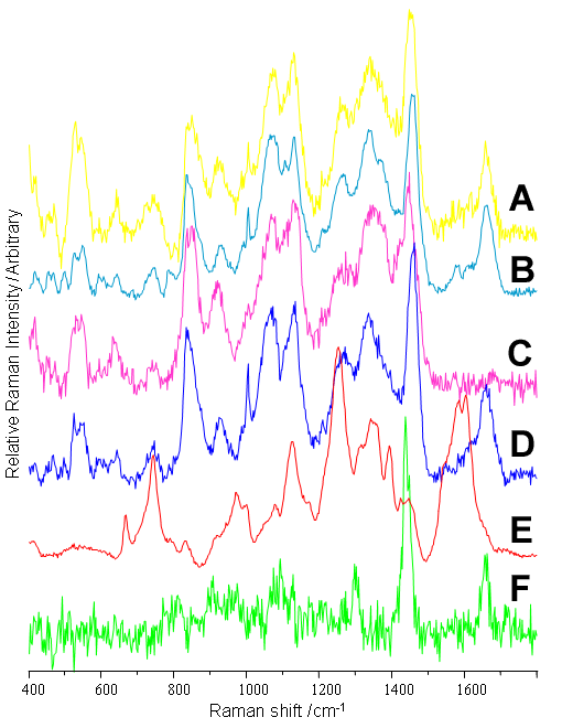
Figure 7. Major Raman signals (633 nm excitation) in the inner layers of porcine retina
Raman spectra, selected from each of the retinal layers were generated using the first significant principal component for that layer. Some components spanned a number of layers; consequently, the total number of spectral contributors mapped was six. A: High t[1] inner nuclear layer (INL; Protein 1), B: Low t[1] INL (DNA), C: High t[1] GCL (Sucrose), D: Low t[1] nerve fiber layer (NFL; Protein 2), E: Low t[1] blood vessel (BLV, Heme), F: High t[1] NFL (lipid). The Raman map in Figure 6 was generated from the linear combination of these reference spectra that accounted for each Raman spectrum. The color coding of the spectral signals is consistent with Figure 6.