![]() Figure 5 of
Beattie, Mol Vis 2007;
13:1106-1113.
Figure 5 of
Beattie, Mol Vis 2007;
13:1106-1113.
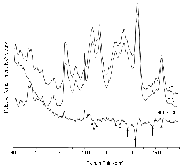
Figure 5. Raman spectra of the nerve fiber layer and ganglion cell layer
The spectra were acquired using 633 nm excitation. Arrows highlight the major bands that decrease on moving from the ganglion cell layer (GCL) to the nerve fiber layer (NFL) are highlighted (arrows). These bands match the differences found between the inner plexiform layer and GCL (Figure 4).