![]() Figure 5 of
Gu, Mol Vis 2007;
13:1045-1057.
Figure 5 of
Gu, Mol Vis 2007;
13:1045-1057.
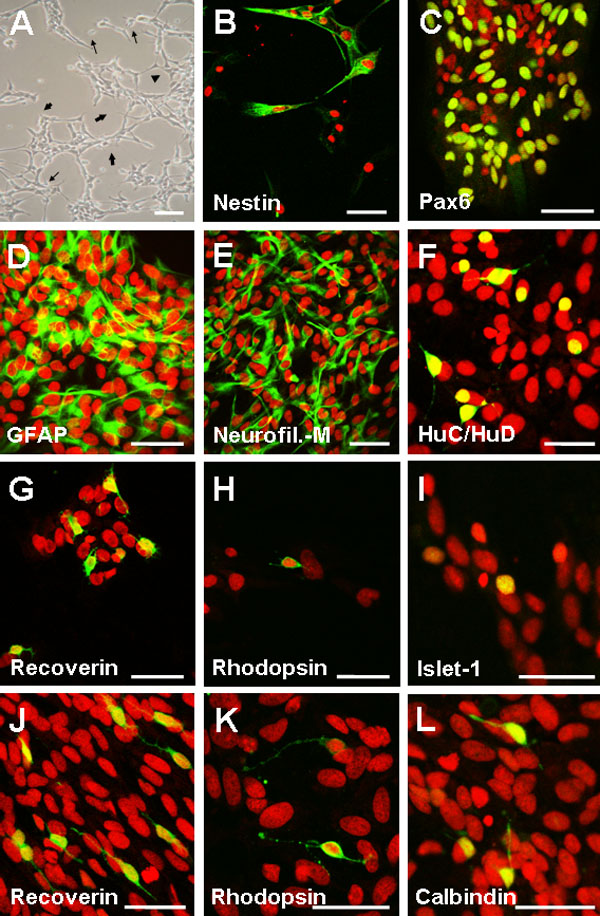
Figure 5. Multipotentiality of retinal stem cells and retinal progenitor cells
Retinal progenitor cells (RPCs) and retinal stem cells (RSCs) were plated on coverslips coated with poly-D-lysine and incubated in differentiation medium for 2 weeks. A: phase contrast microphotograph of serum-treated RPCs showing cells with small bodies and elongated dendritic processes (thin arrows), some apparently connected (thick arrows) as well as cells with large, polygonal shapes (arrowhead) to indicate morphological changes associated with neuronal and glial differentiation. B-I, L: RSCs maintained in differentiation medium for 2 weeks were fixed and immunostained with antibodies to: nestin (B); Pax6 (retinal progenitors, amacrine cells; C); GFAP (glial cells; D); neurofilament-M (RGCs, interneurons; E); HuC/HuD (horizontal, amacrine cells; F); recoverin (cone and rod photoreceptors; G); rhodopsin (rod photoreceptors; H); Islet-1 (bipolar, amacrine cells I); and calbindin (horizontal, amacrine, RGCs; L). J-K: RPCs maintained in differentiated medium for two weeks were fixed and immunostained with antibodies to: recoverin (J), and rhodopsin (K). Cells expressing the same marker differentiated in clusters. Thus, microphotographs of recoverin and rhodopsin immunostaining in RPCs and RSCs are not for quantitative comparison and are not representative of the counts reported in Figure 6. Nuclei were stained with propidium iodide. Scale bars: A, C, and F represents 100 μm; B, D-E, and G-K represents 50 μm.