![]() Figure 1 of
Pang, Mol Vis 2006;
12:756-767.
Figure 1 of
Pang, Mol Vis 2006;
12:756-767.
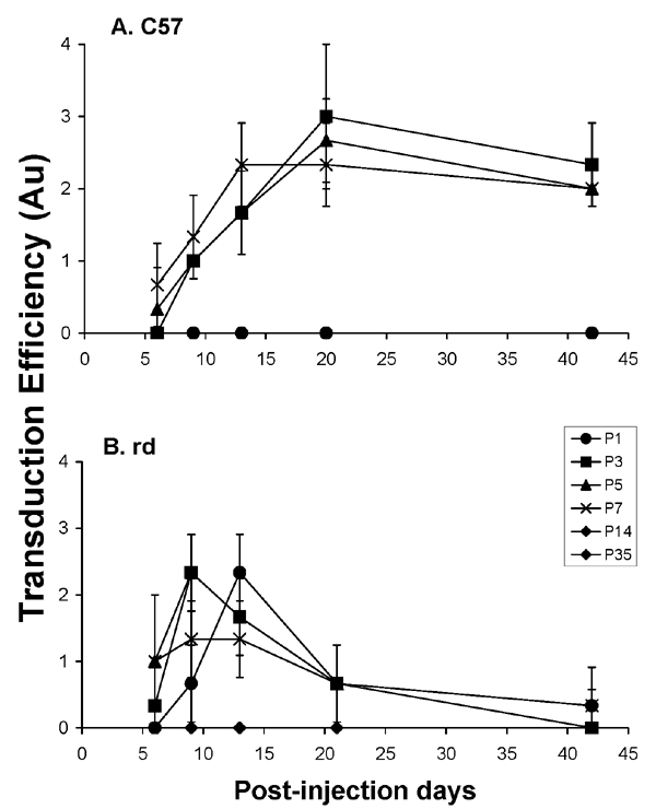
Figure 1. Time course of transduction efficiency following subretinal injections at various ages
The graph shows an analysis of the relationship between the transduction efficiency scores (see Table 1) in normal and rd mouse retinal whole mounts prepared on various days (6 to 42, on the abscissa) after subretinal injections of lenti-GFP that were made at P1, P3, P5, P7, P14, or P35. Transduction efficiency in normal (C57) mice (A) peaked between 13 to 20 days after subretinal injection on P3, P5, and P7 and continued at moderate levels until 42 days post-injection. Virtually no transduction of the vector occurred in the retinas of normal mice injected at P1, P14, or P35. In contrast, when the injection was performed in P1 through P7 rd mice (B), higher transduction efficiency appeared earlier than in the normal mice, but decreased dramatically by 13 to 20 days following injection, the period when all the rods degenerate in this mutant. As in the normal mice, rd mice injected either at P14 or P35 showed no retinal transduction of the vector. Bars indicate the standard deviation. AU represents arbitrary units for transduction efficiency.