![]() Figure 3 of
Pendergrass, Mol Vis 2006;
12:712-724.
Figure 3 of
Pendergrass, Mol Vis 2006;
12:712-724.
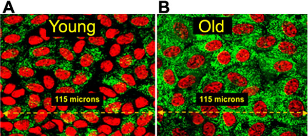
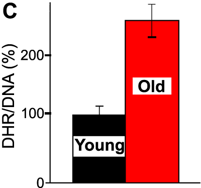
Figure 3.
Higher power views of DHR and DNA fluorescence in nuclei and mitochondria of LECs present on the anterior surfaces of typical young and old rat lenses. This LSCM projection (200x original magnification) shows mitochondria vitally stained with DHR (green) and nuclei vitally stained with Hoechst 33342 (red) of LECs on the anterior surfaces of young (A) and old (B) rat lenses. C: The mean of the DHR/DNA fluorescence ratios (ROS/cell) for the anterior surface epithelia of 11 young compared to 12 old lenses. The error bars represent standard errors of the mean (p<0.001). Other details as described in legend to Figure 1.