![]() Figure 1 of
Pendergrass, Mol Vis 2006;
12:712-724.
Figure 1 of
Pendergrass, Mol Vis 2006;
12:712-724.
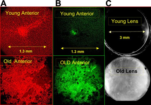
Figure 1.
A comparison of lens anterior surfaces of young and old rat lenses vitally stained for DNA and ROS. A: Comparison of DNA staining with Hoechst 33342 (computer coded red) of the anterior surface of young and old lenses. B: Comparison of vital staining for ROS fluorescence (with DHR shown in green) of the same surface areas. C: Comparison of low magnification photos taken with a reflected light microscope of the anterior sides of the same young and old lenses. Each LSCM image is a projection of the average of 25 separate LSCM frames scanning from the mid anterior surface inwards 100 μm. The confocal settings are identical for young and old pairs so that relative comparisons of DNA fluorescence and cataract reflectance can be made.