![]() Figure 1 of
Allison, Mol Vis 2006;
12:655-672.
Figure 1 of
Allison, Mol Vis 2006;
12:655-672.
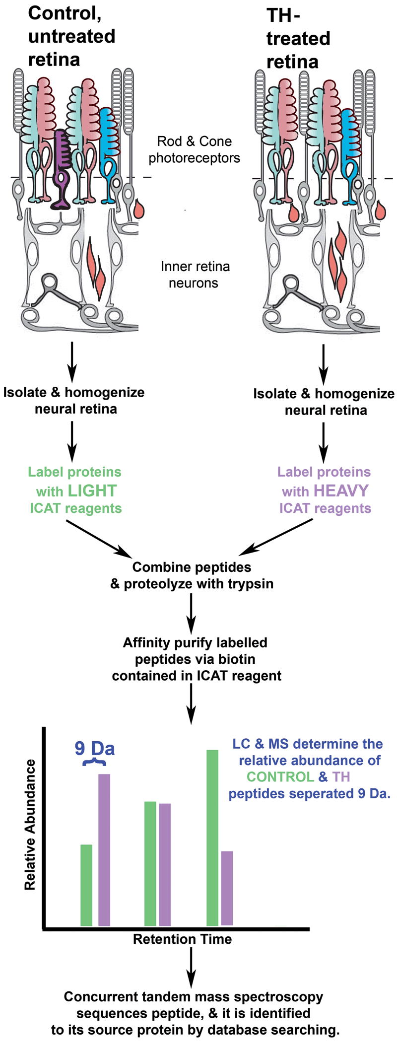
Figure 1. Methodology for identifying peptides and determining their relative abundance
Peptides are isolated from control and thyroid hormone (TH)-treated retinas. Control and treated peptide pools are separately labeled with isotope-coded affinity tag (ICAT) reagents. The ICAT reagents contain biotin, which allows the peptides to be isolated via avidin affinity. The peptides from TH-treated retinas are labeled with ICAT reagents that are 9 Da heavier than the reagents used to label peptides from control retina. The heavy and light isotopes of the ICAT label differ only in that the heavy isotope contains 9 deuterium ions while the light isotope contains hydrogen atoms. This allows the peptides to be identified in mass spectrometry, where their peaks appear 9 Da apart. Identification of these doublets also allows the relative abundance of each peptide to be determined (Figure 2 is an example of this type of spectrum). Concurrently, the peptides are sequenced by mass spectrometry (Figure 2) and searched against protein databases to determine the protein the peptide came from.