![]() Figure 1 of
Steely, Mol Vis 2006;
12:372-383.
Figure 1 of
Steely, Mol Vis 2006;
12:372-383.
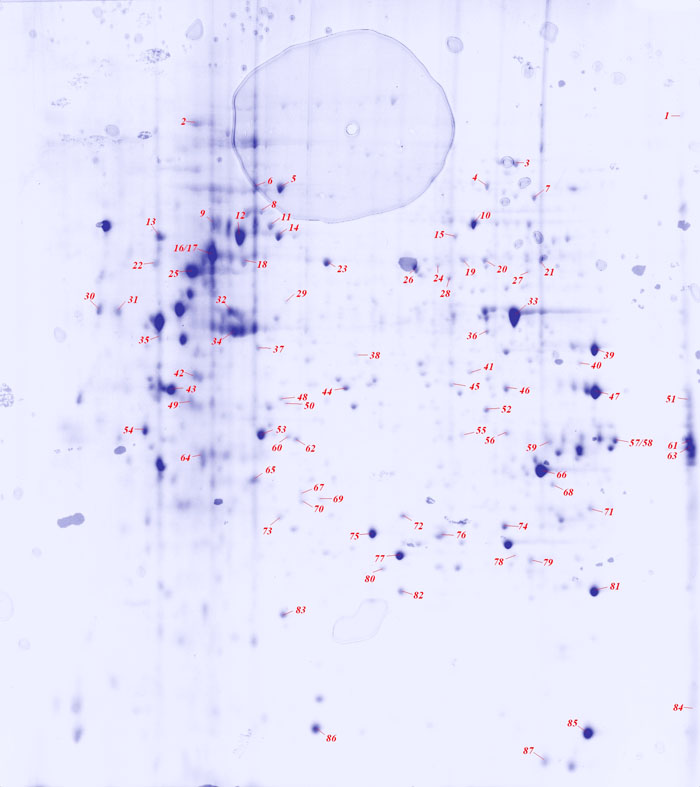
Figure 1. Colloidal Coomassie blue stained two-dimensional gel of GTM3 protein
Image of the Coomassie blue-stained GTM3 gel used to obtain protein spots for identification by mass spectrometry. The spot numbers and lines indicate the gel locations where primary proteins listed in Table 1 were identified.