Figure 3 of Zenteno, Mol Vis 2006; 12:331-335.
Figure 3.
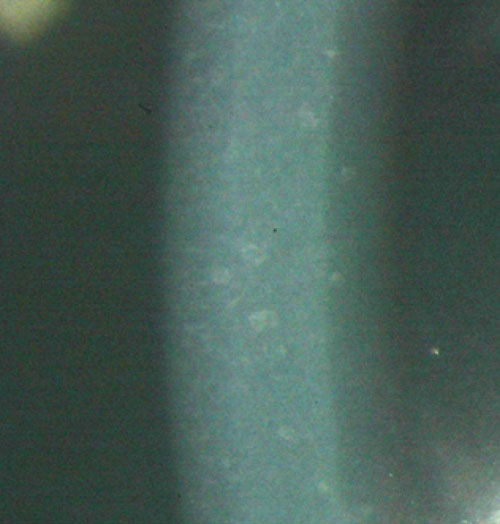
Photograph of the proband's cornea showing granular opacities in the anterior and midstroma. Note that the deposits tend to be more numerous in the anterior corneal stroma.