![]() Figure 1 of
Zenteno, Mol Vis 2006;
12:331-335.
Figure 1 of
Zenteno, Mol Vis 2006;
12:331-335.
Figure 1.
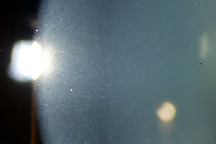
The corneal phenotype of the proband is illustrated by this photograph. Numerous small non-coalescent opacities are evident in the peripheral cornea while a few larger lesions are apparent toward the central part of the cornea.