![]() Figure 9 of
Kuszak, Mol Vis 2006;
12:251-270.
Figure 9 of
Kuszak, Mol Vis 2006;
12:251-270.
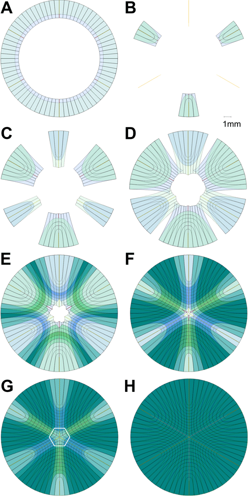
Figure 9. Correct segmental Y suture formation scheme as extrapolated from quantitative analysis of micrographs
By including time, the fourth dimension, in the quantitative analysis of secondary fiber development, animations created with scale CADs reveal novel information about this lifelong process that is not apparent with other analytical methods. Key frames from this animation are shown as static images. This animation begins at the end of fiber rotation and migration, or at the end of fiber middle segment formation (A). At this time it can be seen that posterior fiber elongation (inner blue ring of posterior fiber ends)is slightly ahead (100-150 μm) of anterior elongation (outer ring of anterior end and middle segments). As the animation continues it can be seen that anterior suture branch construction is initiated ahead of posterior suture branch formation (B-D). In point of fact, a short (about 15% of the total branch length) peripheral segment of any anterior suture branch is constructed prior to the initiation of posterior suture formation. D: Essentially, the entire peripheral portion of suture branches are formed before the opposite ends of the same fibers reach their other sutural destination. E: The longest fibers in a growth shell, which are also the fibers with the greatest amount of end curvature, are the last fibers to reach either of their sutural destinations and the first to reach both of their sutural destinations. F,G: Quantification of fiber length reveals that straight fibers are shorter than their immediate neighbors. Thus, solid geometry dictates that the other end of straight fibers that established the origin of posterior suture branches will reach their other sutural destination, the anterior pole, ahead of their neighboring fibers that have opposite end curvature. Similarly, the other end of straight fibers that established the origin of anterior suture branches will reach their other sutural destination, the posterior pole, ahead of their neighboring fibers that have opposite end curvature. As a result, one end of a straight fiber will define a primary, or peripheral origin of a suture branch, while its other end will define a secondary, distal origin of an offset suture branch. The establishment of the secondary, distal origin of suture branches is more readily appreciated by animating the process in larger scale CADs (white hexagonal inset on the forming Y suture in panel G) of the polar caps as seen in Figure 11. This animation ends with the suture patterns fully formed (H) and the fibers having begun the maturation process (indicated by a doubling of a fibers' color value [darkness]). Note that fiber maturation does not occur at the same time in fibers of a growth shell. The different rates appear to be a function of when a fiber reaches both its sutural destinations.
Note that the slide bar at the bottom of the quicktime movie can be used to manually control the flow of the movie. If you are unable to view the movie, a representative frame is included below.
| This animation requires Quicktime 6 or later. Quicktime is available as a free download. |
