![]() Figure 11 of
Kuszak, Mol Vis 2006;
12:251-270.
Figure 11 of
Kuszak, Mol Vis 2006;
12:251-270.
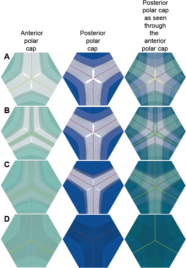
Figure 11. Y suture distal end segment formation
This animation, based on larger scale CADs of the polar region of Y suture lenses, is intended to clarify distal end segment formation. Key frames from this animation are shown as static images. The animation begins with fiber groups made of fewer fibers than in animations 1-5, showing the straight fibers that define the origins of posterior suture branches arriving at the anterior pole ahead of their neighboring fibers and before the straight fibers that define the origins of anterior suture branches arrive at the posterior pole (A). By arriving at the poles ahead of their neighboring fibers with opposite end curvature, formation of a distal end portion of suture branches will be initiated and constructed in a reverse direction (B). The incrementally longer fibers with opposite end curvature on either side of straight fibers that have reached the poles curve away from one another and from the polar axis until they abut and overlap to form the distal portion of suture branches (C,D). Suture branch formation is completed when the forming peripheral and distal portions of suture branches meet within a 10 latitudinal degree circumscribed polar cap.
Note that the slide bar at the bottom of the quicktime movie can be used to manually control the flow of the movie. If you are unable to view the movie, a representative frame is included below.
| This animation requires Quicktime 6 or later. Quicktime is available as a free download. |
