![]() Figure 4 of
Kuszak, Mol Vis 2006;
12:251-270.
Figure 4 of
Kuszak, Mol Vis 2006;
12:251-270.
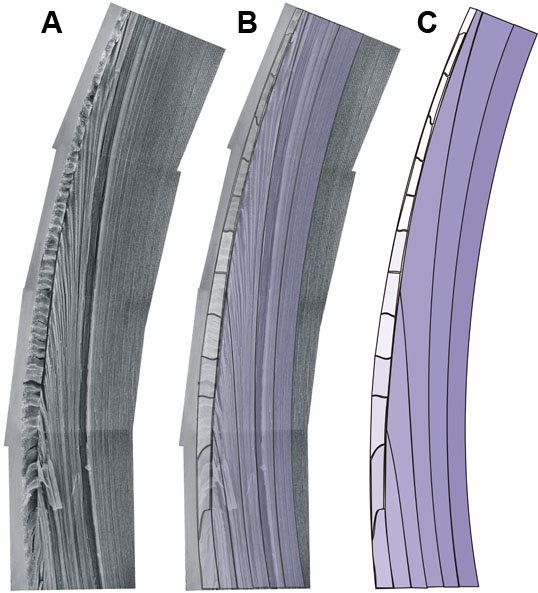
Figure 4. Scale CADs from SEM micrographs showing germinative and transitional zones
A representative portion from a scanning electron micrograph montage of a young adult rat lens radial cell column face showing germinative and transitional zone epithelial cells and portions of nascent fiber cells in the bow region (A). As in Figure 3, the progressive changes in shape, size, orientation, and organization that characterize secondary fibers in radial cell columns are reasonably represented in tracings of groups of fibers (B,C). All of the traced portions of the montage are put together to create a rendition of an entire radial cell column formation.