![]() Figure 9 of
Linberg, Mol Vis 2006;
12:1674-1686.
Figure 9 of
Linberg, Mol Vis 2006;
12:1674-1686.
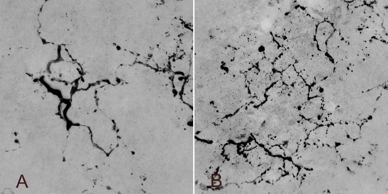
Figure 9. Two HC outgrowths at the retinal surface seen en face
Confocal images of retinal wholemounts of cat retina detached for 28 days taken at the outer retinal surface. The images have been digitally inverted and enhanced using Abode Photoshop. A: Seen en face, two anti-calretinin-positive HC outgrowths gives rise to multibranched and beaded arrays of processes that spread laterally over the retinal surface. While these arborizations appear planar, this "z-series" consists of 20 μm thick optical sections. B: At lower magnification, the multiple arborizations of six outgrowths crowd the retinal surface. Figure 9A is reprinted from Progress in Retinal and Eye Research, 24, S. K. Fisher, G. P. Lewis, K. A. Linberg and M. R. Verardo, "Cellular remodeling in mammalian retina: results from studies of experimental retinal detachment," 395-431, 2006, with permission from Elsevier.