![]() Figure 8 of
Linberg, Mol Vis 2006;
12:1674-1686.
Figure 8 of
Linberg, Mol Vis 2006;
12:1674-1686.
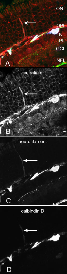
Figure 8. A triple-labeled HC outgrowth seen in each channel
A series of confocal images of the same HC outgrowth from a retina detached for 28 days and triple labeled as described above in Figure 7B. A: A reddish triple-labeled outgrowth rises from processes of the same hue in the outermost outer plexiform layer (OPL). Examining each channel independently demonstrates that this outgrowth is labeled with all three antibody probes. ONL represents outer nuclear layer; INL represents inner nuclear layer; IPL represents inner plexiform layer; GCL represents ganglion cell layer; NFL represents nerve fiber layer. B: The outgrowth labels most intensely with the calretinin antibody that strongly labels both types of HC as well as several types of amacrine cell. C: Antibodies to neurofilament protein strongly label the A-type HC, but also weakly stain the B-type cell. This same weak labeling can be seen in the outgrowth (arrow). Most of the fibers in nerve fiber layer are strongly labeled as are certain ganglion cell processes in the mid-IPL. D: The antibody to calbindin D intensely labels the A-type HC [18], while the B-type, as well as the outgrowth (arrow), are weakly labeled.