![]() Figure 7 of
Linberg, Mol Vis 2006;
12:1674-1686.
Figure 7 of
Linberg, Mol Vis 2006;
12:1674-1686.
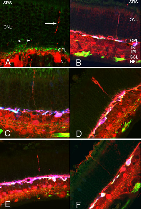
Figure 7. Confocal images of "undirected" HC outgrowths
Confocal images of isolated HC outgrowths most commonly seen in detachments of longer duration. A: Double labeled 7 day detached cat retina. A number of short beaded processes (arrowheads) label with the calretinin antibody (red) and appear directed to retracted rod terminals labeled with the antibody to VAMP2 (green). To the right, a long, unbranching outgrowth (arrow) crosses the outer nuclear layer (ONL) and appears undirected with respect to rod terminals. SRS represents subretinal space; OPL represents outer plexiform layer; IPL represents inner plexiform layer. B: Triple labeled 28 day detached cat retina labeled with antibodies against calretinin (red), neurofilament (green) and calbindin D (blue). A red, predominantly anti-calretinin-positive HC outgrowth rises from red processes in the distal outer plexiform layer (OPL) and crosses the outer nuclear layer (ONL). SRS represents subretinal space; INL represents inner nuclear layer; IPL = inner plexiform layer; GCL represents ganglion cell layer; NFL represents nerve fiber layer. C: 28 day detached retina labeled as in B above and in D to F below. Another red anti-calretinin-positive outgrowth rises through the ONL to the subretinal space. It is unbranched, but has multiple varicosities along its length. D: 7 day detached retina. A red anti-calretinin-positive HC outgrowth rising from the outer OPL appears to broaden as it crosses the ONL. E: 7 day detached retina. Yet another red anti-calretinin-positive HC outgrowth rises into the ONL and has a large varicosity in the inner ONL. F: 7 day detached cat retina. A reddish 'undirected' HC outgrowth rises from the outermost portion of the OPL and passes through the ONL without branching. The ascending fiber maintains a uniform caliber and lacks beads or varicosities. Once at the level of the subretinal space, it gives rise to processes spreading horizontally along the retinal surface. There are no outgrowths rising from the pinkish to white A-type cells. Indeed, in B through F, a plexus, of whitish A-type HC processes courses through the lower OPL without participation in any of these outgrowths. B and F reprinted from Progress in Retinal and Eye Research, 24, S. K. Fisher, G. P. Lewis, K. A. Linberg and M. R. Verardo, "Cellular remodeling in mammalian retina: results from studies of experimental retinal detachment," 395-431, 2006, with permission from Elsevier.