![]() Figure 4 of
Linberg, Mol Vis 2006;
12:1674-1686.
Figure 4 of
Linberg, Mol Vis 2006;
12:1674-1686.
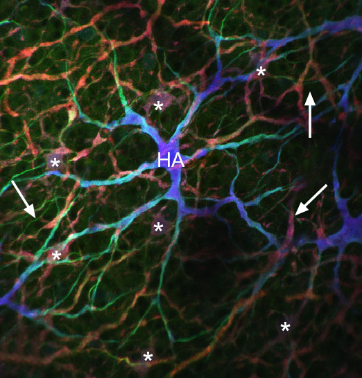
Figure 4. An axonless A type HC in a 7 day wholemounted retina
Wholemount of a 7 day detached retina labeled as in Figure 3. The triple-labeled axonless A type HC (HA) appears bluish-white. The greenish tinge to its distal-most dendrites suggests that more neurofilament protein is expressed there relative to the perikaryon and primary dendrites. Five reddish cell bodies of B-type HCs (*) lie deeper in the INL. Uniformly thin greenish processes (arrows) are likely the axons of the B-type cell showing an upregulation of neurofilament protein in response to retinal detachment. Reprinted from Progress in Retinal and Eye Research, 24, S. K. Fisher, G. P. Lewis, K. A. Linberg and M. R. Verardo, "Cellular remodeling in mammalian retina: results from studies of experimental retinal detachment," 395-431, 2006, with permission from Elsevier.