![]() Figure 10 of
Linberg, Mol Vis 2006;
12:1674-1686.
Figure 10 of
Linberg, Mol Vis 2006;
12:1674-1686.
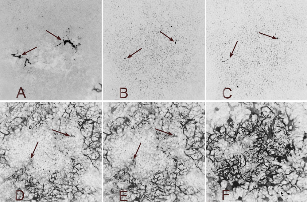
Figure 10. A sequence of confocal images of two HC outgrowths that span the ONL
Images selected from a serial z series through the outer retina. Two HC outgrowths from a wholemount of cat retina detached for 28 days and labeled with an antibody to calretinin. Image processing as in Figure 9. A: The arboreal trunks of the two outgrowths spread laterally at the exposed photoreceptor surface. B: Descending into the outer ONL, the two fibers (arrows) connecting to these outer arborizations can be seen. C: The same two outgrowths (arrows) still deeper in the ONL. One (to the left) runs obliquely in the plane of section. D: At the outermost portion of the OPL, these two fibers connect to their processes of origin (arrows). E: These fibers can be traced for a few sections further (arrows) before being lost in the red tangle of anti-calretinin-positive processes resident to the outer OPL, as seen in F.