![]() Figure 2 of
Suarez, Mol Vis 2006;
12:1467-1472.
Figure 2 of
Suarez, Mol Vis 2006;
12:1467-1472.
Figure 2.
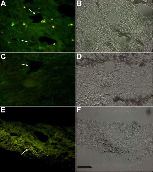
ELAM-1 immunoreactivity in the outflow pathways of porcine and human eyes. A: Immunohistochemical detection of ELAM-1 in the outflow pathway of the porcine eye subjected to experimental glaucoma. Note the fluorescent signal lining the canals and in the trabecular meshwork region (arrows). B: Phase contrast image of the same section presented in A. C: Immunohistochemistry of the outflow pathway of the control porcine eye in which glaucoma was not induced. ELAM-1 immunofluorescence was not observed. D: Phase contrast image of the same section presented in C. E: Detection of ELAM-1 in the human outflow pathway. The section corresponds to a surgical sample obtained from trabeculectomy of a glaucomatous eye. Note the fluorescent labeling at Schlemm's canal and the surrounding trabecular meshwork (arrow). F: Phase contrast image of the section presented in E. Scale bar 25 μm.