![]() Figure 1 of
Suarez, Mol Vis 2006;
12:1467-1472.
Figure 1 of
Suarez, Mol Vis 2006;
12:1467-1472.
Figure 1.
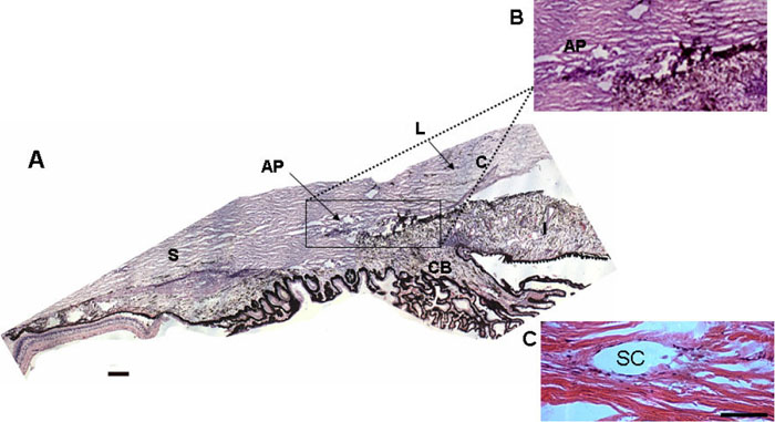
Morphological structure of the porcine and human anterior chamber using hematoxylin&eosin staining. A: Anatomy of the porcine aqueous outflow tract. The section corresponds to an iridocorneal angle. The boxed region marks the drainage outflow system, which is made up of several small canals forming the angular plexus (AP) instead of a single Schlemm's canal found in humans. The aqueous humor produced in the ciliary body (CB) proceeds through the trabecular meshwork into the AP. B: Higher magnification of the aqueous outflow section delimited by the box in A. C: Histological section from a human anterior segment included in the present study. A single Schlemm's canal is illustrated as well as its boundaries. AP, angular plexus; S, sclera; C, cornea; L, limbus; CB, cilliary body; SC, Schlemm's canal. Scale bar 25 μm