![]() Figure 7 of
Tserentsoodol, Mol Vis 2006;
12:1306-1318.
Figure 7 of
Tserentsoodol, Mol Vis 2006;
12:1306-1318.
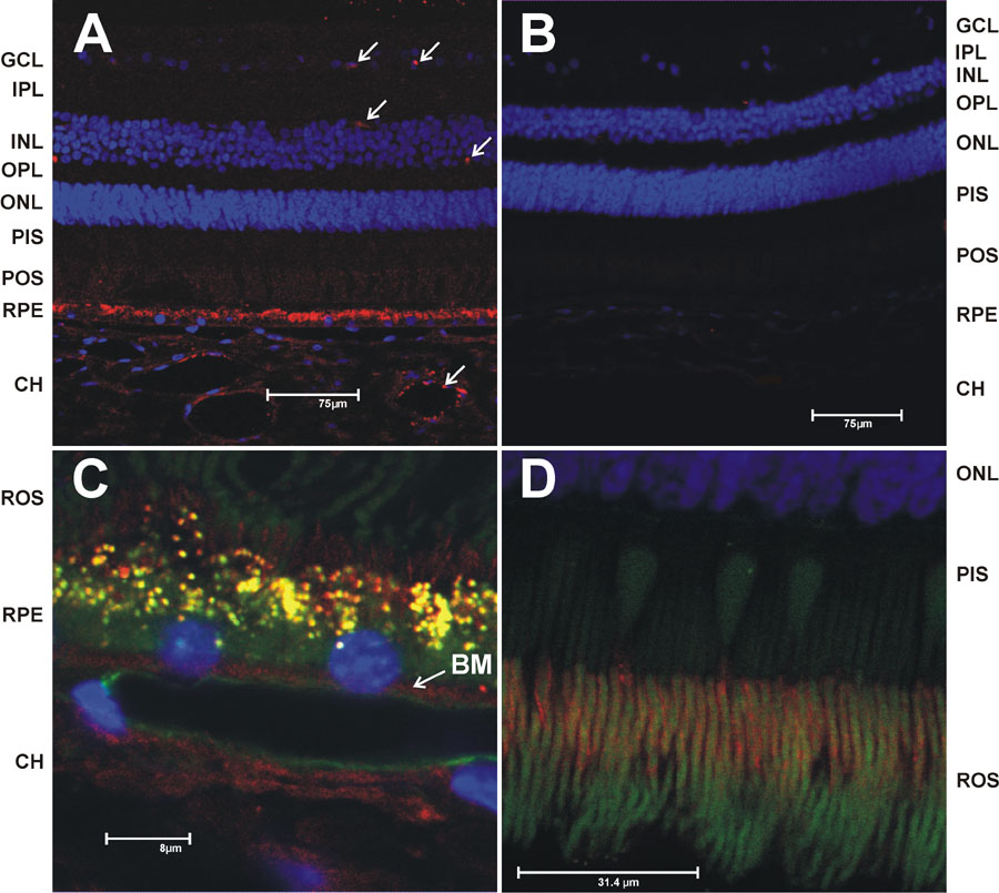
Figure 7. Immunohistochemical localization of apoB
The immunohistochemistry was performed as described in the Figure 6 legend (see also Materials and Methods). A: ApoB immunoreactivity detected with an anti-human apoB100 mouse monoclonal antibody (BD Biosciences) at 1:50 dilution. The top arrows point to immunoreactivity in retina capillaries. The lower arrow in the CH points to LDL deposits in the choroid. B: Negative control (no primary antibody) at same magnification as (A). C: ApoB immunoreactivity at higher magnification focusing on the RPE/CH region. The green channel was added to C and D to allow visualization by autofluorescence of the outer segments and lipofuscin deposits in the RPE. Yellow color in RPE demonstrates co-localization of the lipofuscin deposits (green) and the apoB immunoreactivity (red). The arrow points to punctate apoB immunoreactivity localized to Bruch's membrane (BM). D: Higher magnification of photoreceptor outer segment region with enhanced green channel autofuorecsence to demonstrate slight immunoreactivity in the IPM.