![]() Figure 6 of
Tserentsoodol, Mol Vis 2006;
12:1306-1318.
Figure 6 of
Tserentsoodol, Mol Vis 2006;
12:1306-1318.
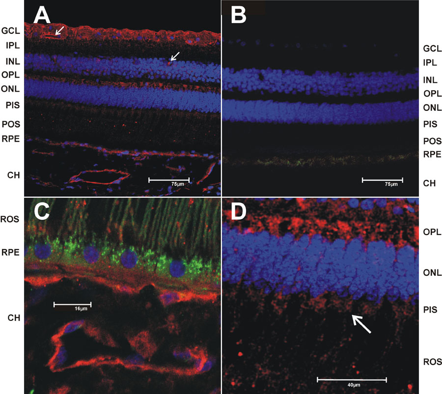
Figure 6. Immunohistochemical localization of low density lipoprotein receptor
Vibrotome sections of monkey retina were immunostained and imaged by confocal microscopy. The nuclei were stained with DAPI (blue), the primary antibodies was detected using Cy5-conjugated (red) secondary antibodies. A: low density lipoprotein receptor (LDLR) immunoreactivity detected with anti-human LDLR rabbit polyclonal antibody (Santa Cruz Biotechnology) at 1:100 dilution. The arrow points to a retinal capillary. B: Negative control (no primary antibody) at same magnification as (A). C: LDLR immunoreactivity at higher magnification focusing on the RPE/CH region. The green channel was added to allow visualization of the rod outer segments and the lipofuscin granules in the RPE by autofluorescence. D: Higher magnification of the photoreceptor inner segments and outer plexiform region (OPL).