![]() Figure 8 of
Colitz, Mol Vis 2006;
12:1067-1076.
Figure 8 of
Colitz, Mol Vis 2006;
12:1067-1076.
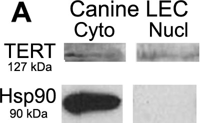
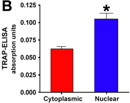
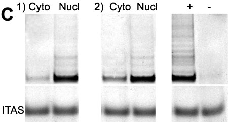
Figure 8.
A: Separation of cytoplasmic and nuclear protein fractions from the first passage of primary cultures of normal canine LEC and western blot analysis for TERT. Both the cytoplasmic fraction (Cyto) and the nuclear fraction (Nucl) show faint expression. Hsp90 was used to assess separation of protein fractions. Cultured LEC lose telomerase activity compared with normal fresh lenses, which may account for the minimal TERT expression by these samples. B: Separation of cytoplasmic and nuclear protein fractions from the first passage of primary cultures of normal canine LEC and evaluation of telomerase activity by TRAP-ELISA; results expressed as absorption units (A). Presence of telomerase activity is defined as being greater than or equal to 0.1000 A. Five separate wells of LEC were separated and evaluated for this experiment, and results are an average of TRAP results from the five separated samples. Results confirm the higher, though still minimal, presence of telomerase activity in the nuclear fraction (0.1054 A) compared to cytoplasmic fraction (0.0628 A), which was negative. This difference was significant (p=0.0012). TRAP-ELISA results for controls are as follows: Positive control (Pos) was 1.837 A, heat inactivated telomerase positive sample served as the negative control (Neg) and was 0.0620 A, and TRS8 (kit control) was 1.389 A. TRS8 is the quantification standard that estimates the amount of product extended by telomerase in a given extract. Each reaction product was amplified in the presence of a 36 bp internal TRAP assay standard (ITAS). C: Separation of cytoplasmic and nuclear protein fractions from the first passage of primary cultures of normal canine LEC and evaluation of telomerase activity by TRAP and polyacrylamide gel electrophoresis. TRAP gels of two of the five samples used in the TRAP-ELISA from Figure 8B are shown in this figure along with the same positive (+) and negative (-) controls. The positive control is provided by the TRAP gel kit and the negative control is a positive sample that has been subjected to heating (85 °C) for 20 min prior to the TRAP assay. The cytoplasmic (Cyto) samples show minimal to no banding while the nuclear (Nucl) samples show faint but obvious banding, consistent with the absorption data.