![]() Figure 2 of
Gendron, Mol Vis 2006;
12:108-116.
Figure 2 of
Gendron, Mol Vis 2006;
12:108-116.
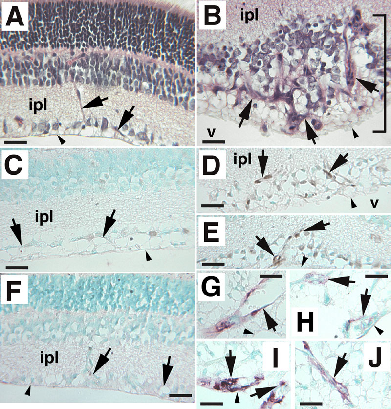
Figure 2. Analysis of blood vessel marker expression in oxygen-induced retinopathy in mice
A: Control normoxia-reared mice showing no retinal pathology. The arrows point to normal retinal blood vessels from a control mouse. B: Hyperoxia-reared mice present large abnormal distended vessels extending along the retina and also exhibit fibrovascular growth. Arrows point to retinal blood vessels, and the bracket indicates a retinal lesion. Nuclear expression of proliferating cell nuclear antigen (PCNA; dense nuclear brown stain, arrows) is upregulated in endothelial cells in cross-sections of retinal blood vessels within the retinal lesions of oxygen-induced animals (D,E), while control animals show low levels of PCNA expression (C). α smooth muscle actin (ASMA) immunostaining (brown reddish staining, arrows) of fibrovascular retinal lesion from oxygen-induced mice (G-J) reveals large abnormal vessels containing myofibroblast cells (arrows) while ASMA expression is not detected in blood vessels of most of the normal control sections analyzed (arrows in F, in which a representative specimen is shown). Low ASMA staining was detected in a total of two retinal blood vessels within three sections of 10 normoxia control retinas analyzed (Figure 4). In all panels, day of analysis is P15, and vitreous body is oriented at bottom of panel. The vitreous (v) and inner plexiform layer (ipl) are identified for orientation. Scale bars represent 25 μm. The retinal inner limiting membrane is indicated by small arrowheads. Original magnification was 250x. Counterstain in panels A,B is hematoxylin and eosin. Counterstain in panels C-J is methyl green. Micrographs shown are representative experiments.