![]() Figure 4 of
Gendron, Mol Vis 2006;
12:108-116.
Figure 4 of
Gendron, Mol Vis 2006;
12:108-116.
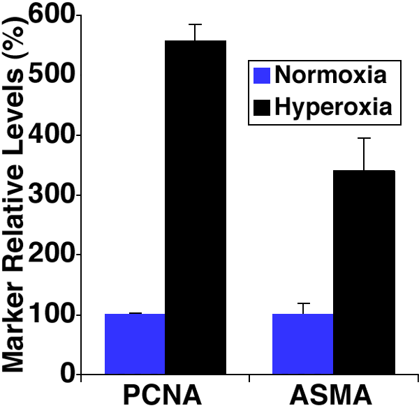
Figure 4. Quantitative analysis of blood vessel marker expression in oxygen-induced retinopathy in mice
Endothelial nuclear expression of proliferating cell nuclear antigen (PCNA) and blood vessel wall expression of α smooth muscle actin (ASMA) in normoxia controls (blue bars) compared to PCNA and ASMA expression in retinal lesions of oxygen-induced animals (black bars) assessed at P15. For PCNA, there were 3 controls and 3 hyperoxic mice. For ASMA, there were 2 controls and 5 hyperoxic mice. Low ASMA staining was detected in a total of two retinal blood vessels within three sections of 10 normoxia control retinas analyzed. Error bars represent SEM. Both PCNA and ASMA expression in hyperoxia-reared were significantly different from control values (p<0.01).