![]() Figure 4 of
Chen, Mol Vis 2006;
12:983-994.
Figure 4 of
Chen, Mol Vis 2006;
12:983-994.
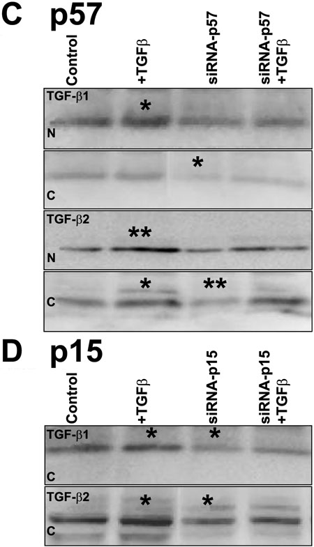
Figure 4.
mRNA and protein analysis of primary cultured human limbal epithelial cells followed by TGF-β treatment after siRNA-p15 and siRNA-p57. A: Relative quantitative real-time PCR analysis showing the effects of siRNA-F and siRNA-p57 on p57 mRNA expression in control and TGF-β (1 ng/ml) stimulated primary cultured human limbal epithelial cells. B: Relative quantitative real-time PCR analysis showing the effects of siRNA-F and siRNA-p15 on p15 mRNA expression in control and TGF-β stimulated (1 ng/ml) primary cultured human limbal epithelial cells. C: Western blot analysis of p57 protein in nuclear (N) and cytoplasmic (C) preparations following TGF-β1 and TGF-β2 treatment (1 ng/ml) for 24 h in primary cultured human limbal epithelial cells with or without siRNA-p57. D: Western blot analysis of p15 protein in cytoplasmic preparation following TGF-β1 and TGF-β2 treatment (1 ng/ml) for 24 h in primary cultured human limbal epithelial cells with or without siRNA-p15. No p15 protein was detected in the nucleus, as shown in Figure 1C. In both C and D, the asterisk indicates a p<0.05 and the double asterisk indicates a p<0.01.