![]() Figure 1 of
Chen, Mol Vis 2006;
12:983-994.
Figure 1 of
Chen, Mol Vis 2006;
12:983-994.
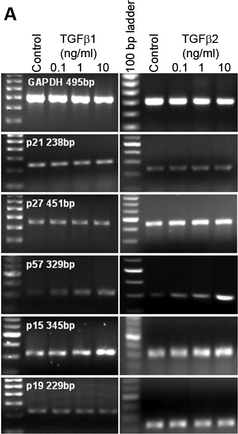
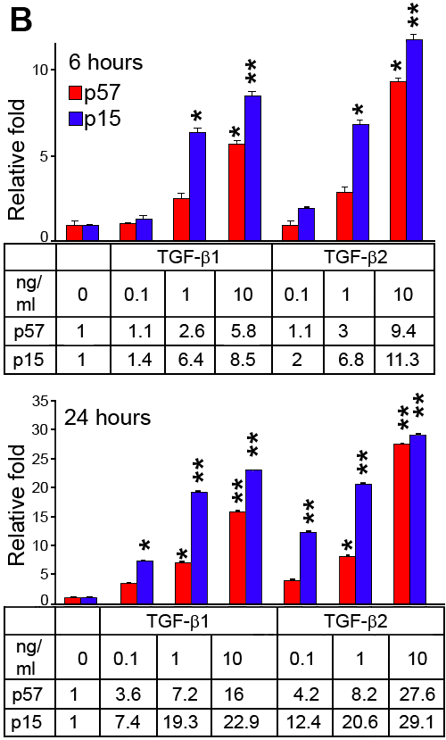
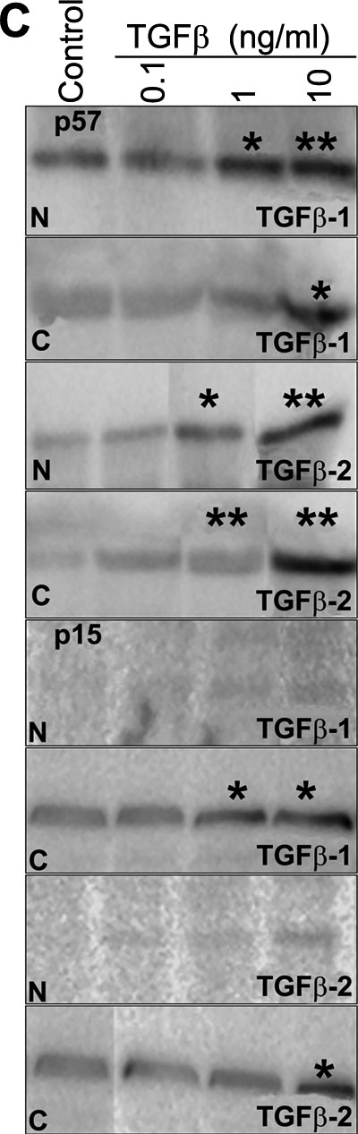
Figure 1.
TGF-β1 and TGF-β2 regulation of cyclin-dependent kinase inhibitors (CDKI) in primary cultured human limbal epithelial cells. A: Semi-quantitative RT-PCR of different doses of TGF-β1 and TGF-β2 treatment for 6 h. B: Relative quantitative real-time PCR for p57 and p15 in primary cultured human limbal epithelial cells before and following TGF-β1 and TGF-β2 treatment for 6 and 24 h. The asterisk indicates a p<0.05 and the double asterisk indicates a p<0.01. C: Western blot analysis showed the location of p57 and p15 before and following TGF-β1 and TGF-β2 treatment of nuclear (N) and cytoplasmic (C) fractions for 24 h. The asterisk indicates a p<0.05 and the double asterisk indicates a p<0.01.