![]() Figure 11 of
Byun, Mol Vis 2006;
12:949-960.
Figure 11 of
Byun, Mol Vis 2006;
12:949-960.
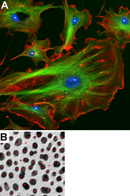
Figure 11. Application of the nuclei detector to different types of images
A: A fluorescent cell image acquired by an epi-fluorescent microscope (downloaded from Image J): actin (red), α-tubulin (green) and nuclei (blue). Six nuclei are detected. B: Blob image (downloaded from Image J). 61 blobs are detected with four detected twice and three false positive blobs.