![]() Figure 10 of
Byun, Mol Vis 2006;
12:949-960.
Figure 10 of
Byun, Mol Vis 2006;
12:949-960.
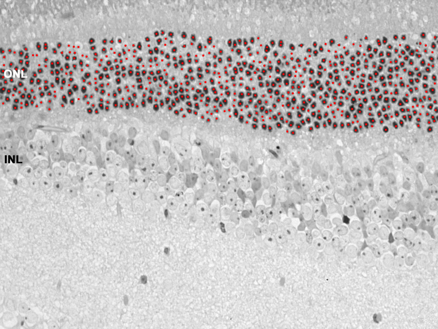
Figure 10. Light micrograph of normal mouse retina
The tissue was embedded in resin and stained with Toluidine blue. The contrast between nuclei and the rest of tissue is low compared to specific nuclear dyes. Red dots on the image represent detected nuclei.