![]() Figure 5 of
Liu, Mol Vis 2006;
12:931-936.
Figure 5 of
Liu, Mol Vis 2006;
12:931-936.
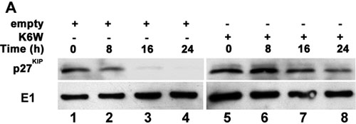
Figure 5.
Overexpression of K6W-ubiquitin inhibits the degradation of p27KIP. HLEC were synchronized and infected with adenovirus as described in Figure 3. After replating, the cells were collected at the indicated times. Levels of p27KIP were determined by western blotting as in Figure 4. The western blot (A) and a densitometry quantification (B) after normalization of protein loading as described in Figure 4 are shown. Experiments were performed three times with reproducible results. The asterisk indicates a p<0.01 as comparing cells infected with adenovirus encoding K6W-ubiquitin with cells infected with empty adenovirus.