![]() Figure 7 of
Whikehart, Mol Vis 2005;
11:816-824.
Figure 7 of
Whikehart, Mol Vis 2005;
11:816-824.
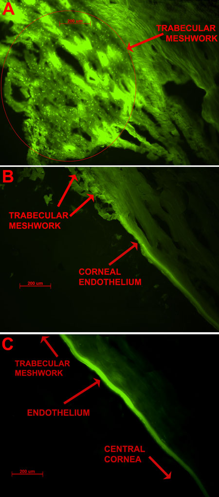
Figure 7. F-BrdU labeling of HCEC, trabecular meshwork, the corneolimbal junction (Schwalbe's line), and the peripheral corneal endothelium after a contact mechanical wound
Human corneal endothelium, trabecular meshwork, the corneolimbal junction (Schwalbe's line), and the peripheral corneal endothelium are shown 48 h after mechanical wounding. A: This image shows the general area of the trabecular meshwork following a contact mechanical wound to the endothelium. Although some cell division was shown here in the unwounded state (Figure 6B), this demonstrates that wounding is accompanied by a comparatively substantial increase in cell division (seen as increased green fluorescence). B: This image indicates that there is significant cell division in the trabecular meshwork, the peripheral corneal endothelium (all indicated by red arrows), and the area between the two anatomical regions (essentially Schwalbe's line). C: The image clearly shows fluorescent labeling in the area of the corneal endothelium (as indicated by the arrow) and suggests that cells in this area are involved in active division by taking up bromodeoxyuridine into new DNA. This panel should be compared with Figure 6B in which essentially no BrdU fluorescence in the control (unwounded) state can be seen. The wound was made in the fellow cornea to that of Figure 6.