![]() Figure 6 of
Whikehart, Mol Vis 2005;
11:816-824.
Figure 6 of
Whikehart, Mol Vis 2005;
11:816-824.
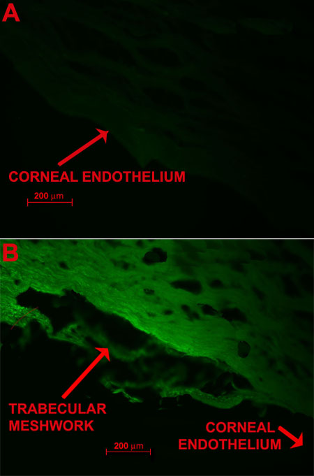
Figure 6. F-BrdU labeling of HCEC and trabecular meshwork in the absence of wounding
A: There is no cell division evident in this non-wounded human corneal endothelium (control). This sample is from a 37-year-old donor which was sequentially exposed to BrdU, primary, and secondary antibodies (the latter coupled to fluorescein). Only a minimal autofluorescent response occurred. The red arrow points to the location of the corneal endothelium. This figure should be compared to Figure 7C in which a wound had been made 48 h prior to fluorescent labeling of the endothelium. The comparison demonstrates how wounding gives evidence of cell division where cell division is normally not present. The BrdU labeling was continued all the way to the central endothelium. B: This image demonstrates the presence of cell division in the human trabecular meshwork under a non wounded (control) condition. The tissue fluorescence is evidence of cell division. Comparing this with Figure 7A suggests that cell division in the trabecular meshwork is substantially increased following wounding.