![]() Figure 2 of
Aldave, Mol Vis 2005;
11:713-716.
Figure 2 of
Aldave, Mol Vis 2005;
11:713-716.
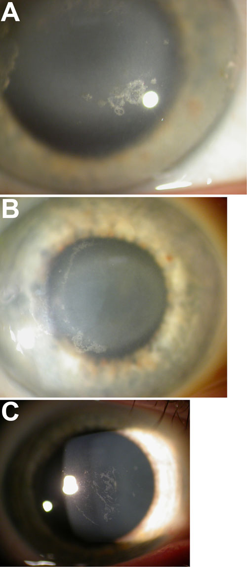
Figure 2. Slit lamp examination of affected from Family 1
A: The 38-year-old proband demonstrating peripheral arcus and annular central corneal opacification with focal, superficial crystalline deposition. B: Peripheral arcus and round central corneal opacification with crescentic crystalline deposition remaining following laser phototherapeutic keratectomy in the proband's 56-year-old mother. C: Irregular, subepithelial, linear and polymorphic opacifications in the proband's 11-year-old son.